Fusion Prostate Biopsy
Fusion prostate biopsy is a type of prostate biopsy that uses advanced imaging technology to improve the accuracy of detecting prostate cancer. A prostate biopsy is a procedure in which small tissue samples are taken from the prostate gland to check for cancer.
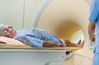
During a fusion prostate biopsy, the patient undergoes a magnetic resonance imaging (MRI) scan of the prostate before the biopsy procedure. The MRI images are then fused with real-time ultrasound images during the biopsy procedure. This allows the healthcare provider to accurately identify and target any suspicious areas within the prostate gland.
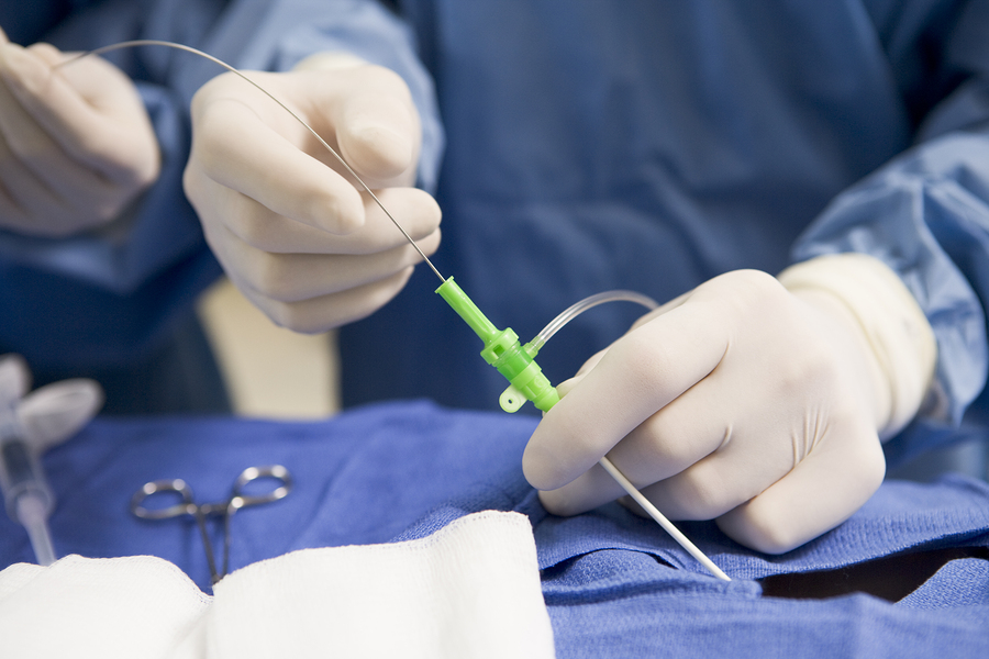
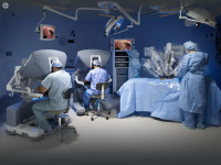
The procedure is performed by a urologist and typically done as an outpatient procedure, under local or spinal anesthesia. During the procedure, the healthcare provider will use a thin, hollow needle to take small tissue samples from the prostate gland. The samples are then examined by a pathologist to check for the presence of cancer cells.
Fusion prostate biopsy is a newer technique that has been shown to increase the detection of clinically significant prostate cancer, particularly in patients with previous negative biopsies or high PSA levels. But it should be noted that this procedure is not suitable for all patients and it should be considered after thorough evaluation and counseling by the healthcare professional.
It's worth mentioning that any biopsy procedure carries a small risk of complications such as bleeding, infection, and pain. Recovery time can vary depending on the patient, but most patients are able to return to normal activities within a few days after the procedure.